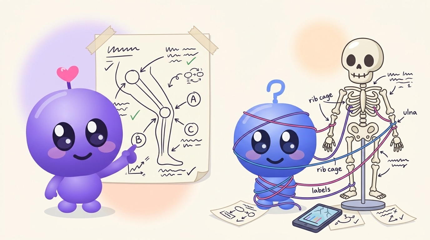
The brachial plexus slide. If you've done a first-year anatomy unit, you know the one: five roots, three trunks, six divisions, three cords, five terminal branches, all crossing over each other in a diagram that looks like a tangled set of earphones. Most students respond by staring at it, copying it neatly, and hoping. A week later it's mush.
Anatomy is easier when you stop treating it as a list of names. Decode each name before you learn it, tie every structure to what it does, and then test yourself on blank diagrams and from memory instead of rereading labelled ones. Do that in short sessions spread across the semester and the names start to hang off a structure you understand, rather than floating around on their own.
Why anatomy feels so heavy
It isn't that anatomy is hard to understand. Most individual facts are simple: this muscle attaches here, this nerve supplies that. The trouble is volume and similarity. There are a lot of structures, many have near-identical names, and the exam asks you to pull the right one out under pressure.
That's a retrieval problem, not an understanding problem. And retrieval is something you can practise directly.
1. Decode the name before you memorise it
Anatomical names are mostly Latin and Greek descriptions. Once you know a few dozen roots, a lot of names explain themselves.
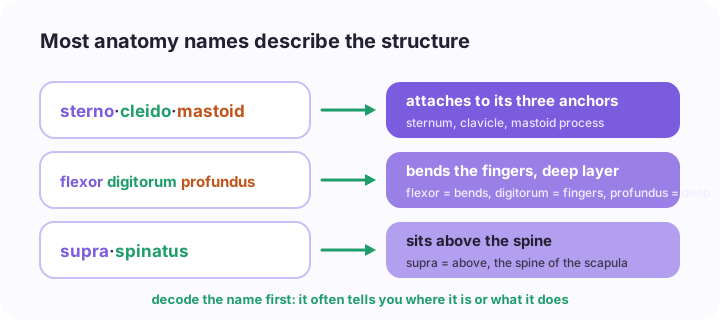
Sternocleidomastoid is a mouthful until you see it's just a list of where it attaches: sternum, clavicle (cleido), mastoid process. Flexor digitorum profundus bends the fingers and sits deep. Supraspinatus sits above the spine of the scapula, and infraspinatus sits below it.
Keep a running page of roots as you meet them: supra and infra, medial and lateral, longus and brevis, profundus and superficialis, the bone names that turn up inside muscle and nerve names. It's the same trick I describe in how to memorise drug names, where the ending tells you the drug class. Learn the parts once and every new name gets cheaper.
Not every name decodes neatly. Some are named after people or are just old. Put those on their own short list and give them extra reps.
2. Learn structure and function together
A muscle's attachments tell you its action. If a muscle crosses the front of the elbow and attaches to the forearm, it's going to help bend the elbow. If you know what a nerve supplies, you can predict what a patient loses when it's damaged.
So for each structure, write the "so what" next to it. For a muscle: attachments, action, nerve supply. For a nerve: where it runs, what it supplies, what goes wrong if it's injured. For a nursing or physio student, that last column is what turns up in clinical questions anyway: which nerve is at risk with a fracture here, why the patient can't do this movement.
Understanding the logic means you can rebuild a fact you've half forgotten. That's worth more than a perfect list you can't reason from.
3. Draw it from memory
Copying a beautiful diagram from your textbook feels productive and teaches you surprisingly little. The useful version is drawing from memory. Close the book, sketch the region roughly (boxes and lines are fine), label everything you can, then open the book and check what you missed.
This is retrieval practice in a form that suits anatomy. Testing yourself beats rereading by a wide margin in learning research, and some smaller anatomy studies suggest drawing helps too, though that evidence is thinner than the case for testing in general. If you want the background, I wrote about it in active recall vs rereading.
For something like the brachial plexus, draw the skeleton of it first (roots, trunks, divisions, cords, branches as columns) before adding names. Once the shape is in your hands, the names have somewhere to go.
4. Quiz yourself on blank diagrams
Every labelled diagram in your lecture slides has a hidden second use. Cover the labels (sticky notes, a screenshot with boxes drawn over them, or just your hand) and name each one. Then flip it: given a name, point to where it is.
Practical exams and spot tests often ask about a structure on a specimen, model or image that looks different from your textbook. So mix up your sources. Look at the same structure in the slides, in an atlas, on a model in the lab and on yourself. Surface anatomy you can feel on your own body (the clavicle, the tibial tuberosity, the pulse points) sticks well because you've touched it.
5. Study by region, revise across systems
Most courses teach anatomy either regionally (the upper limb, then the thorax) or by system (the skeleton, then the muscles, then the nerves). Follow your course for first learning. Then, closer to the exam, revise the other way at least once.
If you learned the upper limb as a region, do a pass where you trace one nerve from the neck to the hand. If you learned by system, do a pass where you name every structure at one level, like a cross section of the forearm. Mixing topics like this feels harder in practice and holds up better in the exam. Interleaving explains why.
6. Little and often beats the Sunday marathon
Anatomy is the textbook case for spaced review. A lot of material, needed for months, much of it reused in later units like physiology and pathophysiology. Fifteen minutes most days, with the structures you got wrong coming back sooner, will hold far more than one long weekend session before the practical.
A rhythm that works for a lot of students:
| When | What | Time |
|---|---|---|
| Same day as the lecture | Decode new names, add roots to your list | 15 min |
| Next day | Draw the region from memory, check and fix | 20 min |
| Most days | Blank-diagram quiz on anything due | 10 to 15 min |
| Weekly | Mixed questions across all regions so far | 30 min |
| Before a practical | Spot-test style: images and models you haven't seen | 30 min a day |
Where The Study Mill fits
I build The Study Mill, so take this with that in mind. If your anatomy unit comes as lecture slides or PDFs, you can upload each week and it writes short lessons, flashcards and a quiz from your lecturer's content specifically, not a generic anatomy syllabus. The quiz mixes multiple choice with written answers that are marked for you, which suits the "explain what happens if this nerve is damaged" style of question. Questions you miss come back in daily review, and Walk & Learn turns the week into audio where Milli and Luke ask you questions out loud, handy on the way to a lab.
What it won't do is replace a good atlas or time with models and specimens. Anatomy is visual and physical, and you need to see and handle structures in three dimensions. Anki is also excellent for anatomy if you're happy to build image occlusion cards yourself. The free plan covers 2 classes, enough to try it on anatomy and one other unit.
Pick next week's anatomy lecture, decode every name on one slide before you try to memorise any of them, then draw the region from memory the following day. You'll find out quickly which bits you know and which ones only felt familiar.